Services
Based on our long experience we are dedicated to providing following radiological services:
MRT
Magnetic resonance imaging
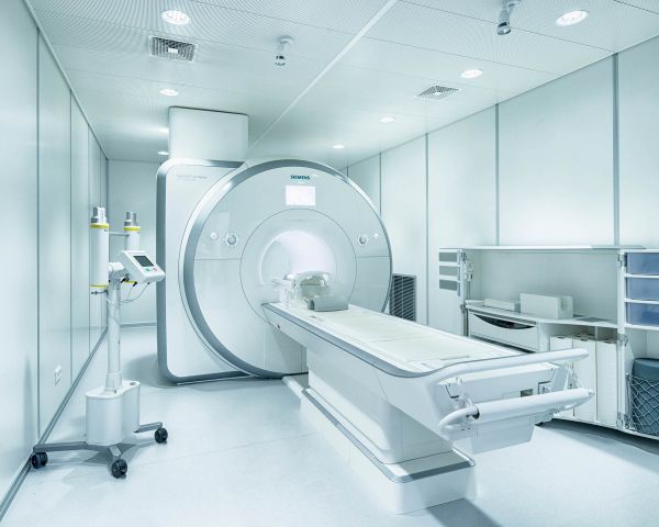
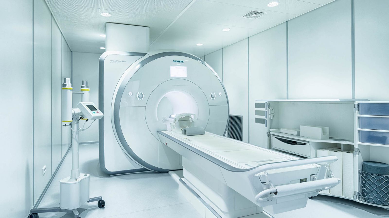
MRI uses a strong magnetic field and radio waves to generate detailed pictures of internal organs and the skeleton in various levels. This technology does not involve X-rays. “Radiologie Ostend” works with MRI machines made by Siemens: Aera and Symphony TIM
From "Head to Toe" the following examinations are provided:
Cervical spine, thoracic spine, lumbar spine, neck organs, chest organs, abdominal cavity organs, gynaecological pelvic, temporomandibular joint and all other joints such as shoulder, knee etc.
Special examinations offered at “Radiologie Ostend” comprise:
Breast MRI, cardiac MRI, MR-Angiography (MRA; imaging of blood vessels).
CT
Computed Tomography
The Spiral-CT used at “Radiologie Ostend” generates numerous sectional images of the body, using X-rays in a continuous spiralling motion. In a computer-based process, these produce multidimensional images.
With our 16 –slice spiral CT scanner made by Siemens, from "Head to Toe" the following examinations can be provided:
All sections of the spinal column, every single joint, neck organs, chest organs and abdominal organs.
Special examinations offered by “Radiologie Ostend” are:
CT Urography (representation of kidneys and the urinary tract), CT Angiography (imaging of blood vessels) , low-dose CT of the thorax and of the paranasal sinuses (NNH).
MAMMO
Digital Mammography
Mammography is a specific type of imaging in which low-dose X-ray radiation is used to examine the breasts. In modern digital mammography, the recordings are electronically processed and archived. Having the capacity of showing changes occurring in the breast before a patient or doctor can even feel them, mammography plays a central role in the early detection of breast cancer.
RÖ
Digital X-ray
Traditional X-ray technology makes use of x-rays which radiate freely through the body. Due to the different intensity of the penetration of individual organs and bones, a print of the object of examination can be generated. Digital X-ray allows images to be electronically processed and archived.
This imaging method is used especially for the thorax and the skeleton, including joints. “Radiologie Ostend’s” digital X-rays are produced with a machine made by “Shimadzu”
SONO
Sonography
Ultrasound, called also sonography, is a method of creating images of the body's interior through the use of high-frequency sound waves. In this type of examination no X-rays are needed. Sound waves are recorded as live images and processed electronically.
With our equipment made by Siemens, “Radiologie Ostend” examines:
abdomen, breast, thyroid, and other soft tissues
Dr. med. Christian-A. Reck
Dr. med. Corinna Possmann
Dr. med. Matthias Tischendorf
Dr. med. Christian-A. Reck
Medical studies at Johann Wolfgang Goethe-University in Frankfurt. Specialist training for radiologist at the municipal hospitals of Darmstadt and Offenbach, completed in 2002. Further specialisation as a neuroradiologist at the University Hospital in Frankfurt, completed in 2005. From 2005 to 2011 head physician at the BG trauma clinic Frankfurt. From 2012 to 2017 head physician of the ZRN Wetterau in Butzbach. Joined our team at "Radiology Ostend" in 2018 as a specialist for neuroradiology and pain treatment under computed tomography.
Dr. med. Corinna Possmann
Medical studies at Johann Wolfgang Goethe-University in Frankfurt and at Humbolt University in Berlin. Medical training at Protestant Geriatric Centre Berlin, Bethanienkrankenhaus Frankfurt, Staedtische Kliniken in Darmstadt and at the German Clinic for Diagnostics in Wiesbaden. Joined our team at “Radiologie Ostend” in 2012 as a specialist for radiology.
Dr. med. Matthias Tischendorf
Radiological training with specialist dental qualification at Friedrich-Schiller-Universitaet Jena, Medical Academy Erfurt and Ruprecht-Karls-Universitaet Heidelberg.


